Ετικέτες

Τετάρτη 9 Μαρτίου 2022

Entwicklung auditiver Verarbeitungs- und Wahrnehmungsleistungen mit und ohne AVWS im Grundschulalter

xlomafota13 shared this article with you from Inoreader

Sprache · Stimme · Gehör 2022; 46: 44-50
DOI: 10.1055/a-1745-7502

Hintergrund Ob sich die an unauffälligen Kindern beschriebenen Reifungsprozesse des zentralen Hörsystems auch an Kindern mit auditiven Verarbeitungs- und Wahrnehmungsstörungen (AVWS) im Grundschulalter nachweisen lassen, sollte an 2 Schülerjahrgängen (1. und 4. Klasse) im Kontrollgruppenvergleich untersucht werden. Material und Methoden In die Auswertung gingen 7 Testergebnisse von 82 Erstklässlern (40 mit AVWS; 42 unauffällige Kinder) und 65 Viertklässlern (35 mit AVWS; 30 unauffällige Kinder) ein. Es wurde eine ANOVA mit dem Gesamtsummenwert aus folgenden 7 Untersuchungen sowie anschließend eine MANOVA mit den Einzeltestergebnissen durchgeführt: Göttinger Sprachaudiometrie II im Störgeräusch; dichotisches Wortpaarverstehen (Uttenweiler-Test); Phonemdifferenzierung, Phonemidentifikation, Phonemanalyse (Subtests aus Heidelberger Lautdifferenzierungstest); Zahlenfolgen-Gedächtnis (Subtest aus psycholinguistischem Entwicklungstest); Mottier-Test. Ergebnisse Die ANOVA zeigte signifikante Haupteffekte von „Schuljahr" (p < 0,001; η² = 0,418) und „Gruppe" (p < 0,001; η² = 0,690), jedoch keine Interaktionseffekte zwischen beiden. Das Ergebnis der MANOVA war ähnlich bzgl. der o. g. Haupteffekte; nur für 2 Tests (Phonemidentifikation, Phonemanalyse) wurde die Interaktion der Faktoren Schuljahr und Gruppe mit jeweils geringen Effektstärken von 3 bzw. 6 % statistisch signifikant. Diskussion Gemäß dieser Querschnittsstudie scheint der Unterschied zwischen den beiden Gruppen unabhängig vom Schuljahr zu sein. Fazit Im Grundschulalter gibt es nicht nur für unauffällige Kinder, sondern auch für solche mit AVWS Hinweise auf eine Reifung des zentralen Hörsystems.
[...]

Georg Thieme Verlag KG Rüdigerstraße 14, 70469 Stuttgart, Germany

Article in Thieme eJournals:
Table of contents  |  Abstract  |  Full text

View on the web

Τρίτη 8 Μαρτίου 2022

A Comprehensive Assessment of Blood Transfusions in Elective Thyroidectomy Based on 180,483 Patients

xlomafota13 shared this article with you from Inoreader

Objectives

To assess the incidence, risk factors, and complications of blood transfusions (BTs) in elective thyroidectomy patients.

Methods

A retrospective cohort study was conducted using the American College of Surgeons National Surgical Quality Improvement Program. Adult patients who underwent elective thyroidectomy from 2005 to 2019 were divided into two cohorts based on whether they received BT or not. Multivariable binary logistic regression models were used to identify risk factors of BT and its impact on postoperative complications.

Results

Of 180,483 patients, 0.13% received BT. Risk factors for BT included underweight body mass index (BMI) (adjusted odds ratio [OR] 3.179, 95% confidence interval [CI] 1.444–6.996), bleeding disorders (OR 2.121, 95% CI 1.149–3.913), anemia (OR 4.730, 95% CI 3.472–6.445), preoperative transfusion (OR 7.230, 95% CI 1.454–35.946), American Society of Anesthesiology physical statuses 3–5 (OR 3.103, 95% CI 2.143–4.492), operative time >150 min (OR 4.390, 95% CI 1.996–9.654), and inpatient thyroidectomy (OR 5.791, 95% CI 3.816–8.787). In addition, transfusion was independently associated with any postoperative complication, non-infectious, cardiac, pulmonary, renal, vascular, or infectious complications, surgical site infection, sepsis, septic shock, wound disruption, pneumonia, unplanned reoperation, prolonged length of stay, and mortality.

Conclusion

Recognition of risk factors of BT is imperative to identify at-risk patients and reduce transfusions by controlling modifiable risk factors such as anemia, operative time, and BMI. In cases where transfusions are still indicated, surgeons should optimize care to prevent or adequately manage transfusion-associated complications.

Level of evidence

3 Laryngoscope, 2022

View on the web

Effect of Hearing Rehabilitation Therapy Program in Hearing Aids Users: A Prospective Randomized Controlled Study

xlomafota13 shared this article with you from Inoreader

Clin Exp Otorhinolaryngol. 2022 Mar 4. doi: 10.21053/ceo.2021.00948. Online ahead of print.

ABSTRACT

BACKGROUND: Despite sufficient hearing gains, many patients with hearing loss have difficulty using hearing aids due to poor word recognition ability. This study was performed to introduce our hearing rehabilitation therapy (HRT) program for hearing aid users and to evaluate its effect on hearing improvement.

STUDY DESIGN: Prospective randomized case-control study.

SUBJECTS AND METHODS: Thirty-seven participants with moderate to moderate-severe sensorineural hearing loss who had used bilateral hearing aids for more than three months with sufficient functional hearing gain were enrolled in this study. Nineteen participants were randomly assigned to the control group (CG) and 18 patients were assigned to participate in our HRT program once a week for eight consecutive weeks (hearing rehabilitation therapy group, HRTG). Their h earing results and questionnaire scores regarding hearing handicap and hearing aid outcomes were prospectively collected and compared between the two groups.

RESULTS: After completing eight weeks of the HRT program, the HRTG showed significantly higher delta score of consonant-only and consonant-vowel sound perception compared to the CG (p<0.05). In addition, the HRTG had a significant improvement in hearing ability as measured by two questionnaires (p<0.05), while no differences were observed in the CG. However, word and sentence recognition test results did not show significant differences between the two groups.

CONCLUSION: Even after short-term HRT, patients had subjectively better hearing outcomes and improved phoneme perception ability; this provides scientific evidence regarding a possible positive role for HRT programs in hearing aid users. Further validation in a larger population with a long-term follow-up study is needed.

PMID:35255665 | DOI:10.21053/ceo.2021.00948

View on the web

Normative measurements of the superior oblique and inferior oblique muscles by magnetic resonance imaging

xlomafota13 shared this article with you from Inoreader

pubmed-meta-image.png

Surg Radiol Anat. 2022 Mar 8. doi: 10.1007/s00276-022-02915-w. Online ahead of print.

ABSTRACT

PURPOSE: Normative oblique muscle data may help to diagnose pathological enlargement of the oblique muscles. We aim to describe the normative values of the superior and inferior oblique muscles in an Australian cohort on T1-weighted MRI and fat suppressed contrast enhanced T1-weighted MRI.

METHODS: A retrospective review of patients who underwent 3 T orbital MRI. The healthy orbits were used to conduct measurements in patients with a unilateral orbital lesion. The maximum diameters of the superior and inferior oblique muscles were measured on coronal planes. The diameter was measured perpendicular to the long axis of the muscles.

RESULTS: The normal measurements (mean ± SD) on fat suppressed contrast enhanced T1-weighted MRI: superior oblique, 3.0 ± 0.5 mm and inferior oblique, 2.7 ± 0.5 mm. On T1-weighted MRI: superior oblique, 2.8 ± 0.5 mm and inferior oblique, 2.5 ± 0.4 mm. In patients who had both sequences performed, the superior and inferior oblique diameters were significantly higher on the fat suppressed contrast-enhanced T1-weighted MRI than the T1-weighted MRI sequence (p < 0.01).

CONCLUSION: Oblique muscle enlargement may be seen in a range of orbital diseases. These data may help in diagnosing oblique muscle enlargement. In addition, variations in the measured muscle diameters can be seen according to the scan sequence th at is used.

PMID:35258651 | DOI:10.1007/s00276-022-02915-w

View on the web

Δευτέρα 7 Μαρτίου 2022

Limited contribution of indocyanine green (ICG) angiography for the detection of parathyroid glands and their vascularization during total thyroidectomy: A STROBE observational study

xlomafota13 shared this article with you from Inoreader

Eur Ann Otorhinolaryngol Head Neck Dis. 2022 Mar 2:S1879-7296(22)00026-6. doi: 10.1016/j.anorl.2022.02.004. Online ahead of print.

ABSTRACT

INTRODUCTION: In total thyroidectomy, indocyanine green (ICG) angiography has mainly been evaluated at end of procedure to predict postoperative hypoparathyroidism. By using it during surgery, we sought to determine whether it could also be an aid to the surgeon.

OBJECTIVE: To determine whether ICG used intraoperatively in total thyroidectomy modified the surgical procedure.

MATERIAL AND METHOD: Thirty-two patients who underwent ICG angiography during total thyroidectomy were included in our single-center retrospective study. The number of parathyroid (PT) glands visualized in white light and on ICG angiography was collected, as well as PT vitality of at end of surgery according to these two modalities. Vitality scores were 0 (no vascularity), 1 (moderately vascularized) or 2 (well vascularize d). Postoperative calcemia at D1, D2 and D7 was analyzed.

RESULTS: In the 32 operations, the surgical procedure was modified in 10 cases (31%). The average number of PTs detected was 2.4 (77 PT) on ICG angiography and 2 (65 PT) in white light. Eleven patients (37.5%) had postoperative hypocalcemia. Cumulative vitality scores at end of procedure were 3.75/8 and 3.37/8 in white light and on ICG angiography respectively (P=0.648). The use of the device did not predict the occurrence of postoperative hypocalcemia.

CONCLUSION: Indocyanine green angiography used in thyroid surgery could assist the surgeon in the identification of PT glands, sparing them in one third of cases.

PMID:35248501 | DOI:10.1016/j.anorl.2022.02.004

View on the web

Current management and perspectives for locally advanced nasopharyngeal carcinoma

xlomafota13 shared this article with you from Inoreader

Cancer Radiother. 2022 Mar 3:S1278-3218(22)00030-0. doi: 10.1016/j.canrad.2021.11.026. Online ahead of print.

ABSTRACT

Nasopharyngeal carcinoma diagnosis is often made at a locally advanced stage (75 to 90% of cases) due to its deep localization. Concomitant radio-chemotherapy is the cornerstone of the treatment of locally advanced forms. The advent of intensity-modulated radiotherapy has improved oncological outcomes and reduced toxicity and is currently the gold standard for irradiation technique. For the locally advanced stage, the addition of induction chemotherapy has become the new standard care according to the latest international recommendations to reduce tumor volumes and act early on micro-metastases. Despite these therapeutic advances, the local and especially distant failure rate remains high. This article reviews current treatment strategies and discuss new approaches and perspectives of locoregional and systemic treatment to re duce treatment failures.

PMID:35249815 | DOI:10.1016/j.canrad.2021.11.026

View on the web

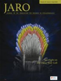
Firing Rate Adaptation of the Human Auditory Nerve Optimizes Neural Signal-to-Noise Ratios

xlomafota13 shared this article with you from Inoreader

10162.jpg

Abstract

Several physiological mechanisms act on the response of the auditory nerve (AN) during acoustic stimulation, resulting in an adjustment in auditory gain. These mechanisms include—but are not limited to—firing rate adaptation, dynamic range adaptation, the middle ear muscle reflex, and the medial olivocochlear reflex. A potential role of these mechanisms is to improve the neural signal-to-noise ratio (SNR) at the output of the AN in real time. This study tested the hypothesis that neural SNRs, inferred from non-invasive assessment of the human AN, improve over the duration of acoustic stimulation. Cochlear potentials were measured in response to a series of six high-level clicks embedded in a series of six lower-level broadband noise bursts. This paradigm elicited a compound action potential (CAP) in response to each click and to the onset of each noise burst. The ratio of CAP amplitudes elicited by each click and noise burst pair (i.e., neural SNR) was tracke d over the six click/noise bursts. The main finding was a rapid (< 24 ms) increase in neural SNR from the first to the second click/noise burst, consistent with a real-time adjustment in the response of the auditory periphery toward improving the SNR of the signal transmitted to the brainstem. Analysis of cochlear microphonic and ear canal sound pressure recordings, as well as the time course for this improvement in neural SNR, supports the conclusion that firing rate adaptation is likely the primary mechanism responsible for improving neural SNR, while dynamic range adaptation, the middle ear muscle reflex, and the medial olivocochlear reflex played a secondary role on the effects observed in this study. Real-time improvements in neural SNR are significant because they may be essential for robust encoding of speech and other relevant stimuli in the presence of background noise.

View on the web

Bioinspired Super-Strong Aqueous Synthetic Tissue Adhesives

xlomafota13 shared this article with you from Inoreader

Matter. 2022 Mar 2;5(3):933-956. doi: 10.1016/j.matt.2021.12.018. Epub 2022 Jan 25.

ABSTRACT

Existing tissue adhesives and sealants are far from satisfactory when applied on wet and dynamic tissues. Herein, we report a strategy for designing biodegradable super-strong aqueous glue (B-Seal) for surgical uses inspired by an English ivy adhesion strategy and a cement particle packing theory. B-Seal is a fast-gelling, super-strong, and elastic adhesive sealant composed of injectable water-borne biodegradable polyurethane (WPU) nanodispersions with mismatched particle sizes and counterions in its A-B formulation. B-Seal showed 24-fold greater burst pressure than DuraSeal®, 138-fold greater T-pull adhesive strength than fibrin glue, and 16-fold greater lap shear strength than fibrin glue. In vivo evaluation on a rat cerebrospinal fluid (CSF) rhinorrhea model and a porcine craniotomy model validated the safety and efficacy of B-Seal for effective C SF leak prevention and dura repair. The plant-inspired adhesion strategy combined with particle packing theory represents a new direction of designing the next-generation wet tissue adhesives for surgeries.

PMID:35252844 | PMC:PMC8896806 | DOI:10.1016/j.matt.2021.12.018

View on the web

Paget's disease of bone and megaloblastic anemia in a 72-year-old patient: A case report and systematic literature review

xlomafota13 shared this article with you from Inoreader

Exp Ther Med. 2022 Apr;23(4):269. doi: 10.3892/etm.2022.11195. Epub 2022 Feb 8.

ABSTRACT

Paget's disease of bone (PDB) is characterized by abnormal osteoclastic bone resorption with disorganized bone neo-formation, primarily affecting elderly (>55 years) patients. Although the majority of patients are asymptomatic, some patients may experience bone pain due to local periosteal involvement or osteoarthritic lesions in the spine; in addition, limb deformities may lead to secondary gait problems or degenerative joint changes. Anemia has an overall prevalence of 12-17% in elderly adults (>65 years old), with macrocytic anemia being the less common type. Megaloblastic anemia is a macrocytic anemia characterized by the presence of large, immature, nucleated cells (megaloblasts) in the blood, with the most common cause being a deficiency of folate and/or vitamin B12. We herein report the rare case of a 72-year-old male patient exhibitin g both these conditions, with the aim of discussing the possible association between the two and, most importantly, the clinical management of the patient in a real-life setting over a period of 10 years. The patient was diagnosed based on clinical symptoms (bone pain), radiological imaging and specific laboratory tests, and received discontinuous courses of bisphosphonates and cyanocobalamin supplementation therapy, based mainly on aggravated symptomatology. A systematic literature review was also performed and revealed not only the scarcity of reports on similar cases, but also the mechanisms that may underlie the possible association of PDB with macrocytic anemia due to vitamin B12 deficiency in elderly patients.

PMID:35251335 | PMC:PMC8892619 | DOI:10.3892/etm.2022.11195

View on the web

A case of musculi peronaeus tertius anatomic variation

xlomafota13 shared this article with you from Inoreader

pubmed-meta-image.png

Surg Radiol Anat. 2022 Mar 7. doi: 10.1007/s00276-022-02899-7. Online ahead of print.

ABSTRACT

In the routine dissections study for medical postgraduates, a rare anatomical variation between the right leg and the ankle was observed on the Asian male cadaver. The peronaeus tertius muscle in this cadaver's right leg divided into two tendons: the first tendon was attached to the base of the fifth metatarsal bone, and the second tendon was inserted into the base of the fourth me tatarsal bone. The purpose of this paper is to provide detailed anatomical case reports, and to discuss the possible causes and mechanisms of the variation by reviewing relevant literature, so as to provide some reference for future anatomical and clinical related disease research.

PMID:35254492 | DOI:10.1007/s00276-022-02899-7

View on the web

Bone fusion in transcele reconstruction of frontoethmoidal meningoencephalocele

xlomafota13 shared this article with you from Inoreader

Br J Neurosurg. 2022 Mar 7:1-5. doi: 10.1080/02688697.2022.2047156. Online ahead of print.

ABSTRACT

OBJECTIVE: In surgical correction of frontoethmoidal encephalocele with transcranial approach, advanced facilities are required. While with extracranial approach, though deemed as a safe option in area with limited facilities, procedure was associated with cerebrospinal fluid (CSF) leakage. In this case series, we evaluate the results of transcele reconstruction of frontoethmoidal encephalocele, our approach to reduce the incidence of CSF leaks by focusing on the closure of layers by its embryological derivatives, by its bone fusion.

METHODS: A case series of 14 patients with various types of frontoethmoidal encephalocele who underwent surgery for defect closure using transcele approach between June 2015 and December 2018 was carried out. Surgery was done by a single surgeon in the Department of Neurosurgery of Cipto Mangunkusumo Hospital in Jakarta, Indonesia. We collected the data of intraoperative blood loss and any signs of infection and CSF leak during the patients' one-year follow up. Bone fusion in the defect was evaluated from 3D rendering of head CT scan that was performed before and in 1 year after surgery.

RESULTS: The median percentage of intraoperative blood loss was 5.9% (0.5-18.7%). All 3D rendering of head CT post-surgery during 1 year follow up showed bone fusion and no patient experienced CSF leaks or CNS infections.

CONCLUSIONS: This study showed that using transcele approach in frontoethmoidal reconstruction could give good bone fusion with minimal blood loss and no CSF leaks. We assumed that closure of the layers by its embryological derivative played an important part in bone fusion and in reducing the incidence of CSF leaks, although this finding has to be validated with large-scale studies.

PMID:35254175 | DOI:10.1080/02688697.2022.2047156

View on the web

Αναζήτηση αυτού του ιστολογίου