Ετικέτες

Κυριακή 31 Ιανουαρίου 2021

Intraparenchymal hemorrhage after surgical decompression of an epencephalon arachnoid cyst: A case report.

paythelady.612 shared this article with you from Inoreader
Related Articles

Intraparenchymal hemorrhage after surgical decompression of an epencephalon arachnoid cyst: A case report.

World J Clin Cases. 2021 Jan 06;9(1):274-277

Authors: Wang XJ

Abstract
BACKGROUND: This study reports the clinical presentation of intraparenchymal hemorrhage as a rare complication after surgical decompression of an intracranial epencephalon arachnoid cyst (IEAC) at the posterior cranial fossa.
CASE SUMMARY: The clinical information of a patient with an IEAC was reported, and the related literature was reviewed. A female patient with nausea presented to our hospital. Computed tomography demonstrated an IEAC located at the posterior cranial fossa, which was large and required surgical intervention. After operation, postoperative intraparenchymal hemorrhage was detected. She had a good recovery with conservative treatment 1 mo later.
CONCLUSION: Though postoperative intraparenchymal hemorrhage is rare after surgical decompression of an IEAC, more attention should be paid to such a complication.

PMID: 33511196 [PubMed]

View on the web

Krukenberg tumor with concomitant ipsilateral hydronephrosis and spermatic cord metastasis in a man: A case report.

paythelady.612 shared this article with you from Inoreader
Related Articles

Krukenberg tumor with concomitant ipsilateral hydronephrosis and spermatic cord metastasis in a man: A case report.

World J Clin Cases. 2021 Jan 06;9(1):278-283

Authors: Tsao SH, Chuang CK

Abstract
BACKGROUND: Tumors of the spermatic cord are rare, and approximately 25% are malignant neoplasms. Metastatic spermatic cord tumors are even rarer. Several studies have revealed that the most frequent primary tumors metastasizing to the spermatic cord and peritesticular tissues are neoplasms of the stomach and prostate. Furthermore, metastasis to the spermatic cord or epididymis may occur via retrograde lymphatic and hematic routes. We present the case of a man with gastric cancer that metastasized to the spermatic cord and epididymis, with concomitant ipsilateral hydronephrosis after surgical resection and chemotherapy for his primary tumor.
CASE SUMMARY: A 71-year-old man underwent total gastrectomy for pT4aN2 poorly differentiated gastric adenocarcinoma in December 2016. Two months after surgery, he received adjuvant chemotherapy with TS-1 from February 2017 to February 2018. Surveillance computed tomography (CT) was performed in June 2018, which did not reveal any sign of tumor recurrence. In November 2019, he presented with left lower quadrant abdominal pain and a palpable left inguinal-scrotal mass. CT revealed left mild hydronephrosis and a left scrotal mass measuring 4.0 cm × 1.7 cm. Tumor biomarkers, including alpha-fetoprotein (AFP), lactate dehydrogenase (LDH), beta-human chorionic gonadotropin (βHCG), carcinoembryonic antigen (CEA), and carbohydrate antigen 19-9 (CA19-9) were all normal. Renal and testicular echography showed left hydronephrosis and a left peritesticular soft tissue lesion with blood flow. Diagnostic ureteroscopy showed left lower ureter narrowing without an intraluminal lesion. A bi opsy was obtained for the indurated spermatic cord and epididymis, which showed poorly differentiated adenocarcinoma. Immunohistochemical staining demonstrated that the tumor was diffusely and strongly positive for homeobox protein CDX2. The features were consistent with metastatic adenocarcinoma of a primary gastric tumor.
CONCLUSION: In patients with a history of primary cancer, an inguinal mass of unknown cause with accompanying ipsilateral hydronephrosis may be a sign of distant metastasis from a primary tumor, especially of gastrointestinal origin.

PMID: 33511197 [PubMed]

View on the web

Simultaneous bilateral acromial base fractures after staged reverse total shoulder arthroplasty: A case report.

paythelady.612 shared this article with you from Inoreader
Related Articles

Simultaneous bilateral acromial base fractures after staged reverse total shoulder arthroplasty: A case report.

World J Clin Cases. 2021 Jan 06;9(1):284-290

Authors: Kim DH, Kim BS, Cho CH

Abstract
BACKGROUND: Acromial and scapular spine fractures after reverse total shoulder arthroplasty (RTSA) are a well-known complication that may negatively impact the effects of long-term outcomes. However, to the best of our knowledge, there has been no report of simultaneous bilateral fractures of the acromion or scapular spine that occurred following staged RTSA.
CASE SUMMARY: A 79-year-old right-handed male visited our outpatient clinic with a chief complaint of pain and limited motion of both shoulder joints for a one-year duration. Based on plain radiographs and magnetic resonance images, the preoperative diagnosis was bilateral cuff tear arthropathy with failed rotator cuff repair. This patient was treated with staged bilateral RTSA at a two-month interval. At 5 and 3 mo after right and left side surgery, the patient returned to the outpatient clinic with severe pain and limited motion of both shoulder joints for 2 wk without a traumatic event. A computed tomography scan revealed non-displaced acromial base fractures of both shoulders. Considering bilateral involvement, fracture location, and patient's demand, open reduction and internal fixation (ORIF) using plate for bilateral acromial base fractures were performed. At 2 years after ORIF, the fracture was completely healed, and the patient was satisfied with shoulder status.
CONCLUSION: This report describes an extremely rare case of simultaneous bilateral acromial base fractures after staged RTSA managed successfully by ORIF with a pre-contoured plate designed for distal clavicle fractures. Although acromial fracture after RTSA can be treated conservatively, simultaneous bilateral fractures may warrant surgical intervention as a means of addressing difficulties in activities of daily living.

PMID: 33511198 [PubMed]

View on the web

Efficacy of cartilage conduction hearing aids in children.

xloma.fota.13 shared this article with you from Inoreader
Related Articles

Efficacy of cartilage conduction hearing aids in children.

Int J Pediatr Otorhinolaryngol. 2021 Jan 16;142:110628

Authors: Nishiyama T, Oishi N, Ogawa K

Abstract
OBJECTIVES: Cartilage conduction hearing aids (CC-HAs) are novel hearing aids that use the third hearing pathway of cartilage conduction. We assessed the efficacy of CC-HAs and the hearing effects and safety of additional tape compression over the transducer in children with hearing loss.
METHODS: The patients (n = 42) underwent a one-month free trial of CC-HAs. Forty of them were patients with auditory canal atresia or stenosis. CC-HA-aided and unaided hearing thresholds (48 fitted ears) were determined using standard audiograms, after which participants could choose to purchase the device or not. We calculated the purchase rates and compared the patient characteristics between the purchase and non-purchase groups along with the purchase reason (or not). We applied additional tape compression over the CC-HA transducer and assessed the hearing effects and side effects.
RESULTS: CC-HA led to hearing improvements at all frequencies. Overall, 72.92% of participants purchased a CC-HA after the trial. By applying additional tape compression over the CC-HA transducer, the stability and hearing gains were improved mainly at low frequencies, and no side effects such as dermatitis were observed.
CONCLUSIONS: CC-HAs are efficacious in producing hearing improvements in children, especially in patients with atresia or canal stenosis who cannot use air conduction hearing aids. Furthermore, we found that the additional tape compression over the transducer was an easy and a safe method for improving the hearing effects and stability of the CC-HA.

PMID: 33494042 [PubMed - as supplied by publisher]

View on the web

Identification of Prostaglandin F2 Receptor Negative Regulator (PTGFRN) as an internalizable target in cancer cells for antibody-drug conjugate development

xloma.fota.13 shared this article with you from Inoreader

journal.pone.0246197.g005&size=inline

by Jorge Marquez, Jianping Dong, Chun Dong, Changsheng Tian, Ginette Serrero

Antibody-drug conjugates (ADC) are effective antibody-based therapeutics for hematopoietic and lymphoid tumors. However, there is need to identify new targets for ADCs, particularly for solid tumors and cancers with unmet needs. From a hybridoma library developed against cancer cells, we selected the mouse monoclonal antibody 33B7, which was able to bind to, and internalize, cancer cell lines. This antibody was used for identification of the target by immunoprecipitation and mass spectrometric analysis, followed by target validation. After target validation, 33B7 binding and target positivity were tested by flow cytometry and western blot analysis in several cancer cell lines. The ability of 33B7 conjugated to saporin to inhibit in vitro proliferation of PTFRN positive cell lines was investigated, as well as the 33B7 ADC in vivo effect on tumor growth in athymic mice. All flow cytometry and in vitro internalization assays were analyzed for statistical significanc e using a Welsh's T-test. Animal studies were analyzed using Two-Way Analysis of Variance (ANOVA) utilizing post-hoc Bonferroni analysis, and/or Mixed Effects analysis. The 33B7 cell surface target was identified as Prostaglandin F2 Receptor Negative Regulator (PTGFRN), a transmembrane protein in the Tetraspanin family. This target was confirmed by showing that PTGFRN-expressing cells bound and internalized 33B7, compared to PTGFRN negative cells. Cells able to bind 33B7 were PTGFRN-positive by Western blot analysis. In vitro treatment PTGFRN-positive cancer cell lines with the 33B7-saporin ADC inhibited their proliferation in a dose-dependent fashion. 33B7 conjugated to saporin was also able to block tumor growth in vivo in mouse xenografts when compared to a control ADC. These findings show that screening antibody libraries for internalizing antibodies in cancer cell lines is a good approach to identify new cancer targets for ADC development. These results suggest PTGFRN is a possible therapeutic target via antibody-based approach for certain cancers.
View on the web

Complicated acute frontal sinusitis in a child presenting with acute ischemic stroke.

xloma.fota.13 shared this article with you from Inoreader
Related Articles

Complicated acute frontal sinusitis in a child presenting with acute ischemic stroke.

Int J Pediatr Otorhinolaryngol. 2021 Jan 22;143:110631

Authors: Davison WL, Gudis DA

Abstract
Acute stroke as a complication of sinusitis is exceedingly rare, and even more so in children. Here we present the case of a healthy 9-year-old male who had an acute stroke in the setting of severe acute pansinusitis. The patient was started on parenteral antibiotics and underwent urgent endoscopic sinus surgery and adenoidectomy, ultimately making a full neurologic recovery. Of the previously reported cases of stroke as a complication of sinusitis, all showed evidence of carotid artery pathology. This is the first report to our knowledge of acute sinusitis presenting as stroke with normal arterial anatomy and function.

PMID: 33508675 [PubMed - as supplied by publisher]

View on the web

Nasopharyngeal microbiome analyses in otitis-prone and otitis-free children.

xloma.fota.13 shared this article with you from Inoreader
Related Articles

Nasopharyngeal microbiome analyses in otitis-prone and otitis-free children.

Int J Pediatr Otorhinolaryngol. 2021 Jan 21;143:110629

Authors: Xu L, Earl J, Bajorski P, Gonzalez E, Pichichero ME

Abstract
OBJECTIVES: About 10-15% children develop frequent acute otitis media (AOM) confirmed by tympanocentesis. These children are designated sOP (stringently defined otitis-prone) because all AOM episodes have been microbiologically confirmed. The cause of otitis-proneness in sOP children is multi-factorial, including frequent otopathogen nasopharyngeal (NP) colonization and deficiency in innate and adaptive immune responses. A largely unexplored contributor to otitis proneness is NP microbiome composition. Since the microbiome modulates otopathogen NP colonization and immune responses, we hypothesized that the NP microbiome composition in sOP children might be dysregulated.
METHODS: We performed 16S rRNA sequencing to analyze microbiome composition in 157 NP samples from 28 sOP and 68 AOM-free children when they were 6 months or 12 months old and healthy. Bioinformatic approaches were employed to examine the composition difference between the two populations and its correlation with changes in levels of inflammatory cytokines.
RESULTS: A different global microbiome profile and reduced alpha diversity was observed in the NP microbiome of sOP children when 6 months old, compared with that from AOM-free children of the same age. This difference was resolved when groups were compared at 12 months old. We found 4 bacterial genera-Bacillus, Veillonella, Gemella, and Prevotella-correlated with higher levels of pro-inflammatory cytokines in the NP. Those 4 bacterial genera were in lower abundance in sOP compared to AOM-free children.
CONCLUSION: Dysbiosis occurs in the NP microbiome of sOP children at an early age even when they were healthy. This dysbiosis correlates with a lower inflammatory state in the NP of these children.

PMID: 33516061 [PubMed - as supplied by publisher]

View on the web

Comparative evaluation of improvement in periodontal and glycemic health status of type 2 diabetes mellitus patients after scaling and root planing with or without adjunctive use of diode laser

xlomafota.13 shared this article with you from Inoreader

Abstract

Background

Periodontal treatment in diabetic patients reduces systemic inflammatory burden and hence should be closely coordinated with the patient's overall clinical diabetic management.

Objective

To evaluate the effectiveness of diode laser (DL) (Biolase EpicTM, 940 nm, Irvine, CA, USA) as an adjunct to scaling root and planing (SRP) on periodontal health and glycated hemoglobin (HbA1c) level of type 2 diabetes mellitus (T2DM) patients suffering from generalized chronic periodontitis (CP), currently, stage II or above/grade B or C periodontitis.

Materials and methodology

After initial screening of 55 T2DM patients, a total of 44 T2DM-CP patients (between the age group of 30 and 65 years) were selected and randomly assigned into two groups. The groups were divided into control group (n=22), treated with scaling and root planing alone (SRP alone), and experimental group (n=22), treated with scaling and root planing along with laser therapy (SRP + DL). Laser irradiation was accomplished at perio pocket setting of 0.8 W (average) in a pulse interval of 1.0 ms and pulse length of 1.0 ms delivering 24 J of energy using a 300-μm fiber optic delivery system.

Results

Thirty-seven out of 44 enrolled T2DM-CP patients completed the study. Both treatment modalities, i.e., SRP alone and SRP+DL resulted in mean significant (p < 0.001) improvement in periodontal health parameters (plaque index (PI), gingival index (GI), probing pocket depth (PPD) and clinical attachment loss (CAL)) and glycemic level (RBS, FBS, and HbA1c) in T2DM-CP patients after 6 months, and was higher in SRP+DL group in comparison to SRP alone. Among the periodontal health parameters, the mean PPD reduction and CAL gain were 51.78% and 48.26% in control as compared to 61.56% and 62.54% in experimental group respectively, whereas the mean significant reduction in HbA1c was 13.8% in SRP alone and 22.52% in SRP+DL group after 6 months (p < 0.05).

Conclusion

Periodontal treatment involving SRP+DL contributes to improved periodontal health parameters and HbA1c level in T2DM-CP patients.

View on the web

Supraclinoid internal carotid artery blister-like aneurysms: hypothesized pathogenesis and microsurgical clipping outcomes

xlomafota.13 shared this article with you from Inoreader

Abstract

Background

Blister-like aneurysms (BLAs) on the supraclinoid segment of the internal carotid artery (ICA) are an enigma of cerebrovascular disease. Neither has a definite pathogenesis been so far identified, nor have uniform treatment guidelines been established for them. Our aim was to develop a hypothesis regarding the evolution of BLAs according to their macroscopic morphologies and to evaluate the efficacy of microsurgical clipping.

Methods

The clinical data and morphological features of 15 consecutive patients with 16 BLAs on the supraclinoid ICA were retrospectively reviewed. The treatment strategies were analyzed, and functional outcomes were evaluated using the modified Rankin scale (mRS). Favorable outcomes were defined as a mRS score of 0–2.

Results

Morphologically, aneurysm growth with expansion of the aneurysm neck before the surgical procedure occurred in two ruptured and one unruptured aneurysm. Daughter bleb formation was observed in two ruptured and five unruptured aneurysms. A varied degree of parent artery sclerosis was observed in nine patients. Thirteen patients were treated with direct surgical clipping, one patient was treated with clipping and wrapping, and the remaining patient was treated with an encircling clipping graft. Favorable and unfavorable outcomes were observed in 13 and two cases, respectively. Follow-up angiograms revealed 4 cases of stenosis with respective degree of mild, 30%, 50%, and 80% without any neurological dysfunction.

Conclusions

We suggest a hypothesis that BLAs on the supraclinoid ICA may share different evolving mechanisms between ruptured and unruptured lesions. A majority of them can be reliably and safely obliterated by direct clipping technique, except for the aneurysms accompanied with severely atherosclerotic parent walls.

View on the web

Πέμπτη 28 Ιανουαρίου 2021

Traumatic brain injury: 10 things you need to know to save lives

Greek DailyNews shared this article with you from Inoreader

Message:

1. THERE'S LITTLE ROOM TO SPARE IN THE CRANIUM
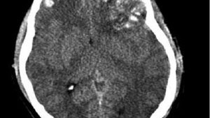
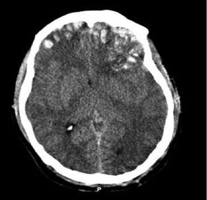
TBI can cause inflammation and swelling or bleeding from damage to brain tissue and blood vessels. Bleeding may occur within the brain (an intracranial hemorrhage) or the protective layers that surround it, such as an epidural, subdural or subarachnoid hemorrhage.

TBI can cause inflammation and swelling or bleeding from damage to brain tissue and blood vessels.
TBI can cause inflammation and swelling or bleeding from damage to brain tissue and blood vessels. (Photo courtesy of Rehman T, Ali R, Tawil I, Yonas H)
RELATED ARTICLES
Stroke: 10 things you need to know to save lives
STEMI: 10 things you need to know to save lives
Trauma: 10 things you need to know to save lives
The brain is tightly enclosed within the skull. Bleeding and inflammation cause a rise in intracranial pressure (ICP), which squeezes and damages brain structures, within that space. As ICP increases, sections of the brain may shift to different areas within the skull, or into the opening where the spinal cord enters the cranium, through a process known as herniation. The higher and longer ICP rises, the degree of permanent disability and likelihood of death increases.

Signs of increased ICP include altered mental status, unequal or nonreactive pupils, posturing, or seizures, as well as bradycardia and hypertension (Cushing's Phenomenon).

2. SLOW DOWN THE DAMAGE FROM SECONDARY BRAIN INJURY
Brain tissue does not regenerate, thus damage from the initial insult is generally permanent. The subsequent edema and bleeding from the primary injury then spreads to damage other areas of the brain, and treatment is aimed at limiting this secondary injury. Through airway protection, oxygenation or ventilation, blood pressure maintenance, and transport to a trauma center, EMS plays a vital role in stopping this cascade.

3. HEAD INJURED PATIENTS ARE UNRELIABLE HISTORIANS
Patients with TBI may be confused, combative, or unresponsive. They may be unable able to follow commands or report pain, and concurrent drug or alcohol intoxication makes assessment even more difficult. Assume that altered mentation is caused by a head injury instead of intoxication.

Rely on inspection, auscultation, and palpation to rule in other injuries, such as a change in the patient's facial expression when their abdomen is palpated. Remember that internal injuries cannot be ruled out, particularly those involving the spinal cord, so take spinal precautions in any patient with a head injury and altered mental status.

4. SOUND THE ALARM AT THE TRAUMA CENTER
Treatment for head-injured patients requires rapid assessment, imaging with a CT scan, and possibly neurosurgery. Time is brain, and the chance of disability or death increases with any delay.

Transporting patients to trauma centers that offer neurological services, and early activation of teams at these centers, streamlines the process to definitive care. Know which hospitals these are, initiate transport as quickly as possible, and alert the hospital to prepare staff and equipment for your arrival.

5. AIRWAY COMPROMISE FROM TBI COMES IN MANY FORMS
Unconscious head-injured patients may lose muscle tone in their jaw, and their tongue may obstruct the airway. The gag reflex may also be compromised in TBI patients, which increases the risk of aspiration from vomit or blood. Clenched teeth, known as trismus, is another common finding that makes position and suctioning airway secretions difficult. Attempt to open the airway with a jaw thrust. Use an oral or nasal airway if the patient will tolerate it, position the patient on their left side to help prevent aspiration, and suction secretions.

Endotracheal intubation is the definitive airway management for TBI, often after sedation and paralysis with RSI. This procedure carries considerable risk, and whether it should be done by ground EMS crews is controversial. Remember that the goal for airway management should be to use the least invasive means to maintain a clear path for oxygenation and ventilation.

6. HYPOXIA AND HYPOTENSION ARE HARMFUL – EVEN FOR A SHORT TIME
Irreversible brain damage can occur in TBI patients after only four minutes of anoxia, which can be caused by a compromised airway, altered respiratory patterns from the head injury, or lung injury in multi-system trauma. Use pulse-oximetry, skin color, and respiratory rate to assess adequate oxygenation. Administer oxygen via nasal cannula, non-rebreather mask, or bag valve to maintain a pulse-ox reading of at least 95%.

Cerebral oxygen delivery is also compromised by hypotension, which is associated with poor outcomes after even transient episodes. Hypotension caused directly by a head injury is a rare and ominous finding; it is more often caused by shock associated with other injuries. Administer IV fluids and titrate blood pressure to 90 – 100 mmHG systolic in head injured patients who are hypotensive.

7. VENTILATING TOO FAST REDUCES BLOOD FLOW TO THE BRAIN
As harmful as hypoxia is in TBI, hypocapnea from excessive ventilation is harmful as well. A higher respiratory rate decreases the amount of CO2 in the blood, which triggers cerebral vasoconstriction and less oxygen delivery. Hyperventilation of head injuries has been associated with poor outcomes.

When assisted ventilation is necessary in isolated head injuries, follow your local protocols regarding use of end-tidal CO2 (EtCO2) monitoring for a head-injured patient.

Shock from multisystem trauma can also cause a low EtCO2 reading, in which case it should not be used to guide ventilation. If capnography is not available, or if the patient may be in shock from other injuries, ventilate adults at 10 breaths per minute. There is one exception...

8. HYPERVENTILATION MAY SLOW HERNIATION
In cases of suspected ICP being so high that portions of the brain may be shifting out of the cranium, the benefit of temporarily reducing cerebral blood with mild hyperventilation may outweigh the harm from less oxygen delivery. Hyperventilation is ONLY indicated for patients who are posturing or have no motor response, have unequal or dilated and nonreactive pupils, or bradycardia and hypotension. Titrate the respiratory rate to maintain an EtCO2 reading between 30 and 35 (in the absence of multi-system trauma), or 20 breaths per minute.

9. YOU MAY HAVE A CONCUSSION
A concussion is a transient change in mentation after blunt head trauma. It can cause retrograde amnesia, and patients often repeatedly ask the same question after being given an answer. It can also cause a temporary loss of consciousness, disorientation, incoherent speech, or lack of coordination. Diagnosis of a concussion is made after a CT scan shows no structural abnormalities associated with these symptoms - not in the field.

While concussion symptoms typically resolve after a few hours or days, severe or multiple concussions can cause headaches, dizziness, depression, and difficulty concentrating for weeks. Especially worrisome is a secondary impact syndrome, where a patient sustains a second concussion before recovering from the first. This can cause rapid cerebral edema and herniation, and has a high mortality rate. Athletes with a head injury should not return to play until after symptoms have completely resolved.

10. BE ON THE LOOKOUT FOR BLOOD THINNERS
Patients taking blood thinners, including Coumadin and Plavix, are at risk of severe bleeding into the brain from relatively minor head injuries. Learn what medications these are, and factor the patient's medication list into your assessment. Signs and symptoms of intracranial bleeding may be delayed, especially in elderly people.

Assume that an altered mental status is a change from baseline unless proven otherwise, and perform a cranial nerve exam to detect subtle signs of closed head injury. Any patient taking a blood thinner who bumps their head should be evaluated at the hospital, no matter how minor their injury appears.

BONUS: AN OUNCE OF PREVENTION CAN SAVE A TON OF HEADACHES
Promoting bike helmet use is one way for EMS agencies to prevent the need to encounter one of the situations listed above. EMS providers are also at risk for head injuries in the back of a moving ambulance. Helmet use is standard in helicopters, and EMS-specific helmets are now available for use on ground ambulances. Consider how easily a concussion may occur from even a minor ambulance crash, and how easily they can be prevented.

Brain_trauma_CT.jpg?w=1600&format=jpg&qu

Proper assessment, treatment, and transport of patients with traumatic brain injury saves lives, here's how
Attachments:
View on the web

Αναζήτηση αυτού του ιστολογίου