Ετικέτες

Πέμπτη 8 Απριλίου 2021

The occult foreign body in the larynx

xlomafota13 shared this article with you from Inoreader
Via hno

pubmed-meta-image.png

HNO. 2021 Apr 8. doi: 10.1007/s00106-021-01043-y. Online ahead of print.

NO ABSTRACT

PMID:33830301 | DOI:10.1007/s00106-021-01043-y

View on the web

Endoscopic Left Central Lymph Node Dissection of Thyroid Cancer: Safe, Feasible, and Relatively Easy

xlomafota13 shared this article with you from Inoreader

pubmed-meta-image.png

Surg Innov. 2021 Apr 8:1553350620983640. doi: 10.1177/1553350620983640. Online ahead of print.

ABSTRACT

Purpose. The purpose of this study was to explore the feasibility of left central lymph node dissection (CLND) in endoscopic thyroidectomy via chest-breast approach (ETCB). Methods. Retrospective analysis of 57 cases of left CLND (group A) via ETCB, 35 cases of open left CLND (group B), and 90 cases of right CLND via ETCB (Group C) were performed from October 2014 t o October 2019. Surgical data, complications, and follow-up data were compared among group A and group B, group A and group C, respectively. Results. There were no significant differences between group A and group B in intraoperative blood loss, tumor size, lymph node (LN) metastasis rate, dissected LN number, metastatic LN number, serum thyroglobulin (sTg), radioactive iodine uptake (RAIU), radioactive technetium uptake (RATU), radionuclide imaging of the residual area (RITRA), and radionuclide imaging of suspicious lymph node metastasis (RISLNM). There were no significant differences between group A and group C in age, operation time, intraoperative blood loss, postoperative hospital stay, tumor size, LN metastasis rate, dissected LN number, metastatic LN number, hypoparathyroidism, sTg, RAIU, RATU, RITRA, and RISLNM. There were 5 cases of temporary recurrent laryngeal nerve (RLN) palsy and 1 case of recurrence in group C. Besides, 1 case of lymphatic leakage was in group A . Conclusion. For selected cases, endoscopic left CLND is safe, feasible, efficient, and more easier than endoscopic right CLND.

PMID:33830818 | DOI:10.1177/1553350620983640

View on the web

Microbiome profiling of uncinate tissue and nasal polyps in patients with chronic rhinosinusitis using swab and tissue biopsy

xlomafota13 shared this article with you from Inoreader

journal.pone.0249688.g005&size=inline

by Sung-Woo Cho, Dong-Young Kim, Sungmi Choi, Sungho Won, Hye-Ryun Kang, Hana Yi

Chronic rhinosinusitis (CRS) is characterized according to the presence or absence of nasal polyps (NPs) and displays nasal microbiota dysbiosis. However, optimal sampling methods of the nasal microbiome in CRS have not been identified. We aimed to assess the microbial composition in patients with CRS, comparing different sampling methods (swab and tissue biopsy), tissue types (uncinate tissue and NP), and disease subtypes. Samples were obtained by swabbing the middle meatus and taking a biopsy of uncinate tissue (UT) in patients with CRS with (CRSwNP, N = 8) or without NP (CRSsNP, N = 6) and controls (N = 8). NPs were also harvested in CRSwNP. DNAs were extracted from fifty-two samples and analyzed by 16S rRNA gene amplicon sequencing. As a result, a great interpersonal variance was observed in nasal swabs, while UT samples presented distinct microbiome with low inter-personal differences. Moreover, the UT microbiomes were further differentiated into three clusters which are associa ted with disease status (control, CRSsNP, and CRSwNP). Compared to UT, NP revealed a unique microbiome profile with significantly less bacterial diversity. Prevotella was the genus whose abundance was negatively correlated with disease severity in NP. In conclusion, tissue samples are better specimens than nasal swabs for assessing the microbiomes of CRS patients. Several bacteria in UT and NP tissues revealed an association with clinical severity of CRSwNP.

View on the web

Spheno-Orbital Meningioma with Cavernous Sinus Extension

xlomafota13 shared this article with you from Inoreader

10-1055-s-0041-1725942_200062ov-1.jpg

J Neurol Surg B Skull Base
DOI: 10.1055/s-0041-1725942

We present a case of a 37-year-old female, with progressive left eye proptosis and an extensive ipsilateral en plaque spheno-orbital meningioma, with diffuse involvement of the lateral wall of the cavernous sinus and the orbit.A cranio-orbital zygomatic approach was performed to reach all extension of the lesion. We made an extradural clinoidectomy and an extensive bone removal of the orbit, exposition of the superior and lateral walls of the cavernous sinus, unroofing of the optic canal, superior orbital fissure, foramen rotundum, and foramen ovale. We performed the peeling of the lateral wall of cavernous sinus with total removal and preservation of the cranial nerv es anatomy and function.In this video, we illustrate the importance of the first time aggressive removal of such tumors. We also demonstrate the concept that secondary invasion of the cavernous sinus are suitable for removal with cranial nerve preservation.The link to the video can be found at https://youtu.be/GJmkqVa6jSs.
[...]

Georg Thieme Verlag KG Rüdigerstraße 14, 70469 Stuttgart, Germany

Article in Thieme eJournals:
Table of contents  |  Abstract  | &nb sp;open access Full text

View on the web

Endolymphatic Sac Tumor Screening and Diagnosis in von Hippel-Lindau Disease: A Consensus Statement

xlomafota13 shared this article with you from Inoreader

10-1055-s-0041-1725033_200331-1.jpg

J Neurol Surg B Skull Base
DOI: 10.1055/s-0041-1725033

Objective Endolymphatic sac tumors (ELSTs) are a frequent cause of hearing loss and other audiovestibular dysfunction in patients with von Hippel-Lindau disease (VHL). Unified screening recommendations for VHL patients have not been established. To develop consensus guidelines, the VHL Alliance formed an expert committee to define evidence-based clinical screening recommendations. Patients and Methods Recommendations were formulated by using the Grading of Recommendations, Assessment, Development, and Evaluation framework after a comprehensive literature review. Results Diagnosis of ELSTs in VHL requires a combination of clinical evaluation and imaging and audiometric findings. Audiovestibular signs/symptoms are often an early feature of small ELSTs, including those that are not visible on imaging. Diagnostic audiograms have the greatest sensitivity for the detection of ELST-associated sensorineural hearing loss and can help confirm clinically relevant lesions, including those that may not be radiographically evident. Magnetic resonance imaging (MRI) can be a more specific test for ELSTs in VHL particularly when supplemented with computed tomography imaging for the identification of small tumors. VHL patients between the ages 10 and 60 years carry high preponderance for ELST presentation. Conclusion We recommend that clinical evaluation (yearly) and diagnostic audiograms (every other year) be the primary screening tools for ELSTs in VHL. We suggest that screening be performed between the ages 11 and 65 years or with the onset of audiovestibular signs/symptoms for synchronicity with other testing regimens in VHL. We recommend that baseline imaging (MRI of the internal auditory canals) can be performed between the ages of 15 and 20 years or after positive screening.
[...]

Georg Thieme Verlag KG Rüdigerstraße 14, 70469 Stuttgart, Germany

Article in Thieme eJournals:
Table of contents  |  Abstract  |  Full text

View on the web

Combined Transcavernous and Kawase's Approach for Gross Total Resection of a Cavernous Sinus Meningioma

xlomafota13 shared this article with you from Inoreader

10-1055-s-0041-1725025_200157ov-1.jpg

J Neurol Surg B Skull Base
DOI: 10.1055/s-0041-1725025

Microsurgery of cavernous sinus (CS) lesions is generally considered to be associated with a high rate of morbidity and cranial nerve deficits. The success for surgical removal of CS meningiomas is debatable and achieving a good functional outcome with preservation of the cranial nerves is the goal. Surgery of these lesions is challenging, recurrence rates are high, and therapeutic strategies remain controversial. In this video, we present a case of a CS meningioma that extended to Meckel's cave and the posterior fossa in a 46-year-old woman with history of a left-sided cerebellopontine angle World Health Organization (WHO) grade-I meningioma with extension to the lef t CS. Seven years ago, she had a microsurgical resection of a Cerebellopontine angle (CPA) meningioma. She later received radiotherapy for the slowly growing meningioma of the posterior CS. The patient presented with newly onset headache and facial pain. Magnetic resonance imaging (MRI) showed a meningioma of the left CS and Meckel's cave extending into the ambient cistern, with a mild mass effect on the pons, and a size increase compared with prior imaging. Since this area previously received radiotherapy, and the patient was symptomatic from trigeminal compression, the decision was made to proceed with surgical resection of the tumor via combined transcavernous and anterior petrosectomy. Postoperatively, the patient woke up with the same neurological status. MRI confirmed gross total resection of the tumor. The histopathology was a WHO grade-II chordoid meningioma. The patient is currently receiving radiotherapy. This video demonstrates the surgical approach and the resection step s of this pathology.The link to the video can be found at: https://youtu.be/HrU1VOzUGWU.
[...]

Georg Thieme Verlag KG Rüdigerstraße 14, 70469 Stuttgart, Germany

Article in Thieme eJournals:
Table of contents  |  Abstract  |  open access Full text

View on the web

Pretemporal Transcavernous Approach for Resection of an Orbito-Cavernous Oculomotor Schwannoma

xlomafota13 shared this article with you from Inoreader

10-1055-s-0041-1724116_200354ov-1.jpg

J Neurol Surg B Skull Base
DOI: 10.1055/s-0041-1724116

The following operative video demonstrates surgical tenets and nuances of the pretemporal transcavernous approach in an unusual case of a 33-year-old patient suffering new onset diplopia and a third nerve palsy due to an orbito-cavernous oculomotor schwannoma. Near total resection was accomplished through an extended pterional craniotomy with pretemporal transcavernous exposure of her lesion, resulting in resolution of her preoperative oculomotor palsy and visual dysfunction. When combined with extended pterional and modified frontotemporal orbitozygomatic approaches, the pretemporal transcavernous approach provides excellent surgical access to the parasellar region i ncluding the superior orbital fissure and cavernous sinus. Meticulous dissection and early identification of tissue planes, including cranial nerve and vascular anatomy, allows for safe removal of tumors arising in this region.The link to the video can be found at: https://youtu.be/EuIRTP7wWBQ.
[...]

Georg Thieme Verlag KG Rüdigerstraße 14, 70469 Stuttgart, Germany

Article in Thieme eJournals:
Table of contents  |  Abstract  |  open access Full text

View on the web

Clip Reconstruction of a Previously Leaked Large Complex Posterior Communicating Segment Aneurysm with Extradural Anterior Clinoidectomy

xlomafota13 shared this article with you from Inoreader

10-1055-s-0041-1726425_200159ov-1.jpg

J Neurol Surg B Skull Base
DOI: 10.1055/s-0041-1726425

Fusiform or near-fusiform aneurysms that involve the long segment of the supraclinoid internal carotid artery (ICA) pose significant challenges to neurovascular surgeons. Involvement of the origin of vital branching arteries in this segment may preclude safe treatment with flow diverting stents. In addition, clip reconstruction may also not be possible in this region due to entire or near-entire involvement of the circumference of the ICA (Fig. 1). In this video article, we present a case of a complex and previously leaked, (visualized with hemosiderin) aneurysm of the posterior communicating segment of the ICA, in a 60-year-old female. Multiple complexities made this aneurysm challenging to treat. These included (1) a 270-degree encirclement of the ICA with multiple lobulations that left only a small section of nondiseased vessel wall, (2) a relatively short segment of the supraclinoidal ICA that made proximal control challenging thus requiring an extradural anterior clinoidectomy, (3) a fetal posterior communicating artery that originated immediately proximal to the beginning of the aneurysm, and lastly, (4) an anterior choroidal artery that was firmly adherent over the aneurysm dome. In this video, we present the microsurgical steps for dealing with this complex aneurysm, including extradural clinoidectomy and clip reconstruction (Fig. 2). Postoperatively, the patient woke up without any deficits. Angiography showed complete obliteration of the aneurysm.The link to the video can be found at: https://youtu.be/C3Vc0maWChc.
[...]

Georg Thieme Verlag KG Rüdigerstraße 14, 70469 Stuttgart, Germany

Article in Thieme eJournals:
Table of contents  |  Abstract  |  open access Full text

View on the web

Τρίτη 6 Απριλίου 2021

Man With Fever and Groin Mass

xloma.fota.13 shared this article with you from Inoreader
A 54-year-old man with a history of hypertension, hyperuricemia, and dyslipidemia presented to the emergency department (ED) with fever and a mass in the left side of the groin since 3 days earlier. One month before presenting to the ED, he received a diagnosis of a urinary tract infection and was treated with oral antibiotics. On arrival in the ED, he was well oriented and had a temperature of 38.1°C and 100.58 °F. Physical examination revealed a firm, tender, longitudinal mass over the left side of the groin (Figure 1).
View on the web

High prevalence of methotrexate use in patients with Epstein–Barr virus-positive mucocutaneous ulcer may cause confounding bias

xloma.fota.13 shared this article with you from Inoreader

Modern Pathology, Published online: 23 March 2021; doi:10.1038/s41379-021-00798-7

High prevalence of methotrexate use in patients with Epstein–Barr virus-positive mucocutaneous ulcer may cause confounding bias
View on the web

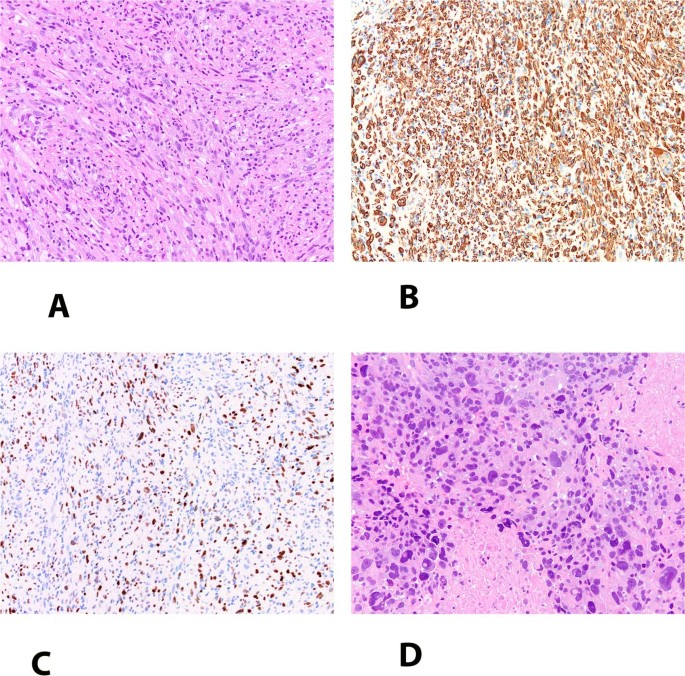
Inflammatory rhabdomyoblastic tumor with progression to high-grade rhabdomyosarcoma

xloma.fota.13 shared this article with you from Inoreader

41379_2021_791_Fig1_HTML.jpg

Modern Pathology, Published online: 23 March 2021; doi:10.1038/s41379-021-00791-0

Inflammatory rhabdomyoblastic tumor with progression to high-grade rhabdomyosarcoma
View on the web

Αναζήτηση αυτού του ιστολογίου